-
Address:
17888 67th Court North
Loxahatchee, FL
-
Mail us:
contact@wrightacademia.org
- submit manuscript
Case Report |
Open Access |
Volume 1 | Issue 1 |
A Rare Case of a Giant Retroperitoneal Lipoma with Multiple Limb and Trunk Lipomata without Familial Multiple Lipomatosis
Jason Russell Laurens, MBBS1 , Adam John Frankel, BSc, MBBS (Hons), PhD, FRACS, FRCSEd1, Bernard Mark Smithers, AM, MBBS, FRACS, FRCSEng, FRCSEd2,3, Geoffrey Strutton4
1Upper Gastro-intestinal and Soft Tissue Unit, Princess Alexandra Hospital, Australia
2Mayne Professor and Head, Discipline of Surgery, The University of Queensland, Australia
3Director, Upper Gastro-intestinal and Soft Tissue Unit, Princess Alexandra Hospital, Australia
4Department of Anatomical Pathology, Princess Alexandra Hospital, Australia
*Corresponding author: Jason Russell Laurens, MBBS, Department of Surgical Specialties, Upper Gastro-intestinal and Soft Tissue Unit, Princess Alexandra Hospital, Woolloongabba, QLD 4102, Australia, Tel: 0414-745-913
Citation: Laurens JR, Frankel AJ, Smithers BM, StruttonG (2021) A Rare Case of a Giant Retroperitoneal Lipoma with Multiple Limb and Trunk Lipomata without Familial Multiple Lipomatosis. J Surg Clin Rpts 1:002.
Copyright © Laurens JR, et al.
Received: |
Accepted: |
Published: |
Introduction
Retroperitoneal lipoma are exceedingly rare, and due to the difficulty in distinguishing between retroperitoneal lipoma and well-differentiated liposarcoma, the treatment recommendation is en-bloc resection. We report the rare and unusual case of giant retroperitoneal lipoma in association with multiple limb and trunk lipoma.
Case Report
A 58-year-old male presented to his general practitioner with right scrotal swelling that developed over a week. He had an ultrasound followed by a computed-tomography (CT) scan, which demonstrated a right hydrocoele, but also showed a well-defined lipomatous mass occupying much of the left side of the lower abdomen, extending from the edge of Gerota's fascia behind the inguinal ligament towards the lesser trochanter. It was posterolateral to the external iliac artery and vein as they exited the pelvis. There was mild dilatation of the left upper ureter likely due to mass effect, but the left pararenal fat did not appear involved in the mass (Figure 1). Subtle heterogeneity was noted on the scans which prompted a provisional diagnosis of well-differentiated liposarcoma (WDLS).
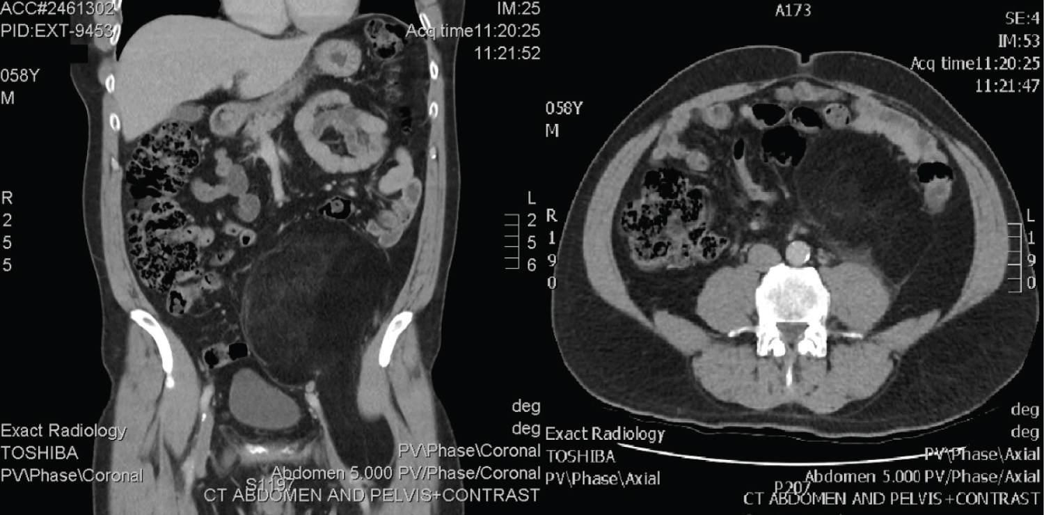
Figure 1: CT showing retroperitoneal mass.
He was referred to our sarcoma service. Medical history was dyslipidaemia, four coronary artery stents for ischaemic heart disease, an unprovoked deep vein thrombosis in his left leg 13 years previously, transurethral resection of the prostate for benign prostatic hyperplasia and lumbar discectomy. He was an active smoker (15/day). His medications were clopidogrel, rosuvastatin and perindopril. There was no family history of lipomas. The abdominal mass was easily palpated in his left lower quadrant and had a soft consistency. There was a hydrocele in the right hemiscrotum. Notably, he had approximately ten subcutaneous soft, well-circumscribed mobile lipomata up to 5 cm on his trunk and limbs. Full blood picture, including renal and liver function tests were normal.
Following discussion in the sarcoma multi-disciplinary team (MDT) meeting, en-bloc resection was recommended. At laparotomy, a large left-sided retroperitoneal mass was found, macroscopically it appeared to be dark yellow to orange adipose tissue within a semitranslucent capsule, involving the psoas and displacing the L2 trunk (Figure 2). Macroscopically it did not involve Gerota's fascia or the mesocolon, allowing kidney and bowel preservation. The left external iliac and common femoral vessels were not involved, and the left femoral nerve was stretched on the anterolateral surface and able to be preserved. A separate incision in the groin was required with division of the inguinal ligament to resect the mass where it was adherent to the psoas insertion at the lesser trochanter (Figure 3). The tumour was removed en-bloc. A Jaboulay procedure of the right hydrocele was performed. He was discharge post-operative day 10, as he was mobilising independently, he was not prescribed chemical DVT prophylaxis. Hispostoperative course was complicated by a superficial thrombosis of the left leg treated with low molecular weight heparin and graduated compression stocking. Although he had a previous unprovoked left leg DVT, and new left leg superficial thrombus, no prothrombotic workup was undertaken due to this thrombus having an identified risk factor, reduced mobility, and bed rest. At six-month review, he had fully recovered.
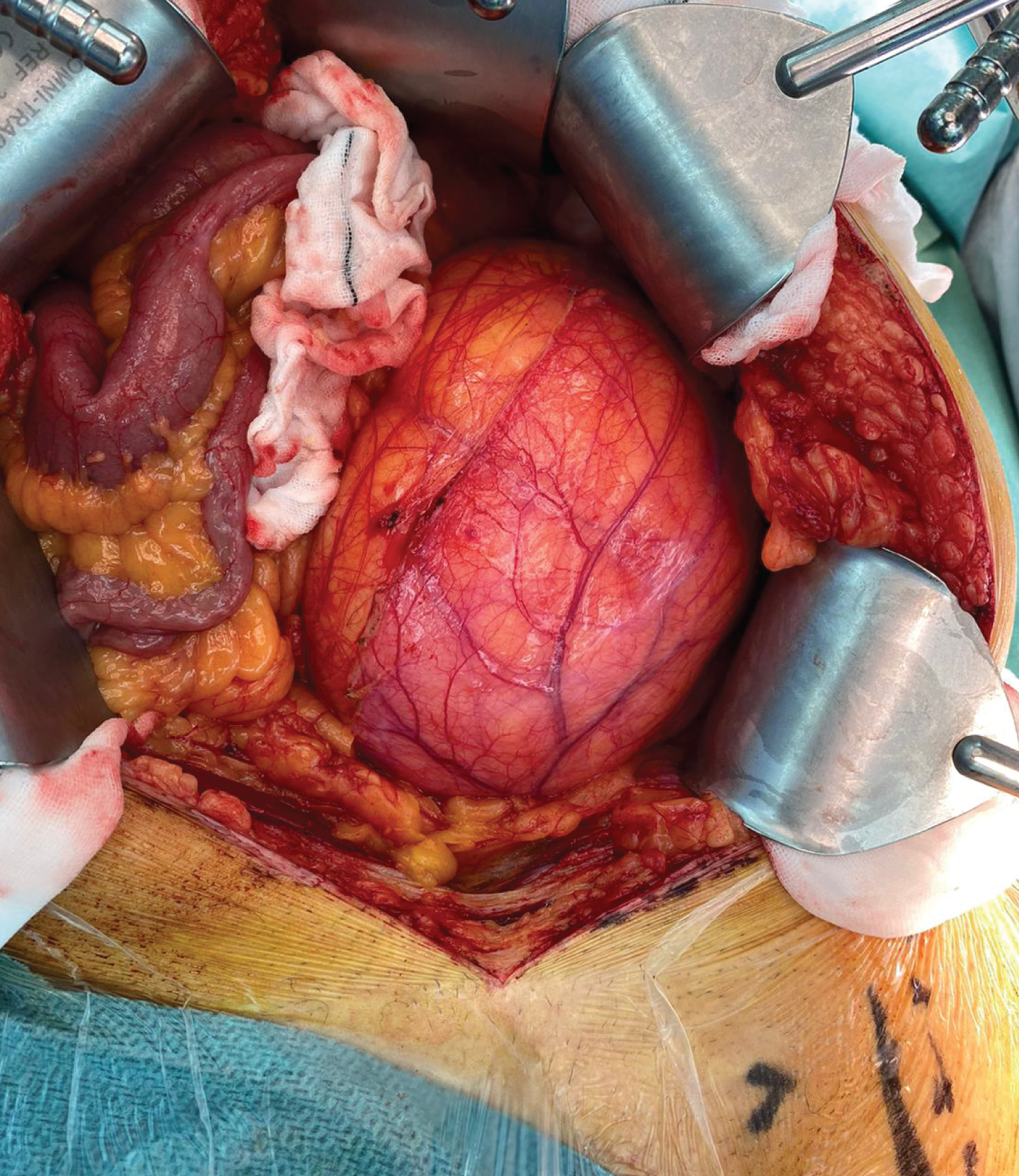
Figure 2: Intra-operative photo showing size of lesion and displacement of bowel.
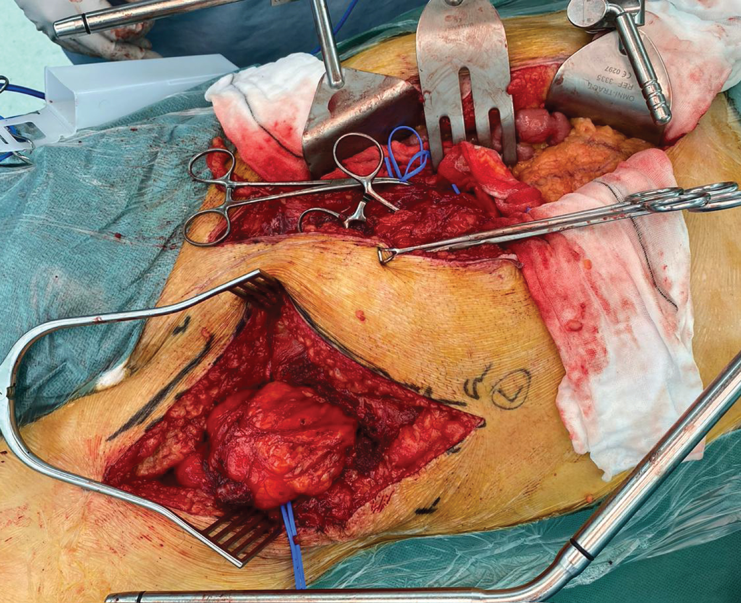
Figure 3: Intra-operative photo showing groin incision with inguinal ligament divided, to allow adequate dissection in lower pelvis and groin.
Histology reported a 160 mm × 150 mm × 90 mm fatty tumour weighing 1540g. Microscopically, an expert soft tissue pathologist favoured lipoma. MDM2 gene amplification was not present on fluorescence in situ hybridisation (FISH) following testing of multiple sites of the tumour. He underwent whole exome sequencing (WES) to further investigate HMGA2 with review at the Queensland Molecular Tumour Board. No significant somatic signatures were identified.
Discussion
Giant retroperitoneal lipomas are remarkably rare, with 20 cases reported in the English literature [1]. While there is no consensus which distinguishes giant lipomas from non-giant lipoma, all previously reported cases of giant retroperitoneal lipoma describe tumours with at least one dimension greater than 10 cm [1]. Fatty tumours of the retroperitoneum represent a diagnostic dilemma, due to the difficulty in distinguishing between benign lipoma and liposarcoma, particularly WDLS [2]. CT and MR imaging cannot definitively diagnose benign or malignant adipocytic lesions [3], radiological features of retroperitoneal lipoma include fat signal attenuation and contain few if any septations while WDLS also demonstrate fat attenuation and inversely commonly contain septa [4]. WDLS often but not always contain mature fatty elements or non-adipose tissue [4,5]. While the Retroperitoneal Sarcoma Transatlantic Working Group recommend image-guided percutaneous core biopsy [6], it is important to acknowledge the accuracy of sampling via core biopsy has been recorded to be 85% for WDLS [7,8]. No studies report core sampling of retroperitoneal lipoma, and there is clearly potential for sampling error with such large tumours. Positive amplification of the MDM2 gene supports a diagnosis of WDLS, however, a negative biopsy does not exclude the diagnosis due to varied amplification among different cells in the same tumour [9]. En bloc resection is the cornerstone of management, which applies equally to WDLS and to large, radiographically 'benign' lipomatous masses, although the preservation of specific organs should be considered on an individual basis [6]. Due to the rarity of giant retroperitoneal lipoma, and the presence of multiple limb lipomata, HMGA2 gene testing was undertaken in order to assess for familial multiple lipomatosis, which is a rare disease characterised by multiple lipomas of the trunk and limbs. Its underlying genetic cause is unknown, although literature suggests deregulation of the MHGA2 gene which encodes for aberrant cell proliferation and development of benign tumours may be responsible [10]. Despite him having multiple superficial lipomas, his testing was negative.
Authors Declarations
No grants or financial assistance was provided for this case report.
All authors are in agreement with the content of the manuscript.
Written informed consent was obtained from the patient for publication of this case report and accompanying images
References
- Chen YH, Chen MJ, Lin HJ (2019) Giant retroperitoneal lipoma presenting as abdominal protrusion: One case report and review of literature. Adv Dig Med 6: 128-131.
- Bibi M, Rhouma SB, Ouanes Y, et al. (2018) Fatty tumors of the retroperitoneum: Lipoma or well-differentiated liposarcoma. About a case of giant retroperitoneal liposarcoma. Urol Case Rep 21: 58-60.
- Morosi C, Stacchiotti S, Marchiano A, et al. (2014) Correlation between radiological assessment and histopathological diagnosis in retroperitoneal tumors: analysis of 291 consecutive patients at a tertiary reference sarcoma center. Eur J Surg Oncol 40: 1662-1670.
- Craig WD, Fanburg-Smith JC, Henry LR, et al. (2009) Fat-containing lesions of the retroperitoneum: Radiologic-pathologic correlation. Radiographics 25: 69-85.
- Francis IR, Cohan RH, Varma DG, et al. (2005) Retroperitoneal sarcomas. Cancer Imaging 5: 89-94.
- Trans-Atlantic RPS Working Group. (2015) Management of Primary Retroperitoneal Sarcoma (RPS) in the Adult: A Consensus Approach from the Trans-Atlantic RPS Working Group. Ann Surg Oncol 22: 256-263.
- Young R, Snow H, Hendry S, et al. (2020) Correlation between percutaneous biopsy and final histopathology for retroperitoneal sarcoma: A single‐centre study. ANZ Journal of Surgery 90: 497-502.
- Ikoma N, Torres KE, Somaiah N, et al. (2015) Accuracy of preoperative percutaneous biopsy for the diagnosis of retroperitoneal liposarcoma subtypes. Ann Surg Oncol 22: 1068-1072.
- Jacob E, Erickson-Johnson MR, Wang X, et al. (2006) Assessment of MDM2 amplification using fluorescence in situ hybridization on paraffin-embedded tissue discriminates atypical lipomatous tumors from lipomas. Mod Pathol 19: 13A.
- Mejia Granados DM, de Baptista MB, Bonadia LC, et al. (2020) Clinical and Molecular Investigation of Familial Multiple Lipomatosis: Variants in the HMGA2 Gene. Clin Cosmet Investig Dermatol 13: 1-10.